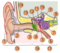
Fitxer:Anatomy of the Human Ear blank.svg

Mida d'aquesta previsualització PNG del fitxer SVG: 659 × 518 píxels. Altres resolucions: 305 × 240 píxels | 611 × 480 píxels | 977 × 768 píxels | 1.280 × 1.006 píxels | 2.560 × 2.012 píxels.
Fitxer original (fitxer SVG, nominalment 659 × 518 píxels, mida del fitxer: 59 Ko)
Historial del fitxer
Cliqueu una data/hora per veure el fitxer tal com era aleshores.
| Data/hora | Miniatura | Dimensions | Usuari/a | Comentari | |
|---|---|---|---|---|---|
| actual | 18:53, 12 abr 2019 |  | 659 × 518 (59 Ko) | Mikael Häggström | Removed misleading green area: The pinna is also part of the outer ear |
| 17:42, 10 set 2018 |  | 659 × 518 (61 Ko) | Jmarchn | Bigger (proportional real size) and full redraw (more realistic) of the auricle. Ossicles in white colour. Eardrum with contour. Added 3 labels. Add fundus to the bone and subcutaneous tissues, add superior auricular muscle, add transparency to middle ear, add separation between middle and inner ear, add division to internal auditory canal. | |
| 14:15, 16 set 2009 |  | 730 × 556 (71 Ko) | M.Komorniczak | {{Information |Description={{en|1=A diagram of the anatomy of the human ear.}} {{pl|Schemat budowy ucha ludzkiego.}} |Source=*File:Anatomy_of_the_Human_Ear.svg |Date=2009-09-16 12:14 (UTC) |Author=*File:Anatomy_of_the_Human_Ear.svg: Chittka L, |
Ús del fitxer
La pàgina següent utilitza aquest fitxer:
Ús global del fitxer
Utilització d'aquest fitxer en altres wikis:
- Utilització a pl.wikipedia.org
- Kosteczki słuchowe
- Młoteczek
- Strzemiączko
- Ucho
- Szablon:Ucho
- Trąbka słuchowa
- Kowadełko
- Błona bębenkowa
- Ślimak (anatomia)
- Ucho zewnętrzne
- Kanały półkoliste
- Małżowina uszna
- Przewód słuchowy zewnętrzny
- Narząd Cortiego
- Śródchłonka
- Błędnik błoniasty
- Ucho środkowe
- Jama bębenkowa
- Błędnik kostny
- Przychłonka
- Łagiewka (anatomia)
- Woreczek
- Mięsień napinacz błony bębenkowej
- Mięsień strzemiączkowy
- Musculus fixator stapedis
- Staw kowadełkowo-młoteczkowy
- Schody przedsionka
- Schody bębenka
- Jama sutkowa
- Przewody półkoliste
- Wodociąg przedsionka
- Przedsionek ucha wewnętrznego
- Przewód śródchłonki
- Worek śródchłonki
- Płatek ludzkiej małżowiny usznej
- Okienko przedsionka
- Okienko ślimaka
- Wikipedysta:Paweł Ziemian/Navi/Ucho
- Wikipedysta:Paweł Ziemian/Navi/Ucho2
- Utilització a pl.wiktionary.org
- Utilització a rm.wikipedia.org


